Development of a multimodal anthropomorphic liver phantom for navigated tumor treatment
Liver tumor resection is a challenging intervention for the surgeon, especially in laparoscopic procedures where hidden lesions cannot be localized through palpation. Within these procedures, the accurate localization of the main vessel branches in relation to the lesions position, is a crucial step to obtain negative resection margins while maintaining sufficient healthy tissue.
Intraoperative ultrasound, in combination with surgical navigation can provide superior orientation by showing the position of surgical instruments together with a registered preoperative image (e.g. MR/CT). Whilst ultrasound provides intraoperative imaging of the underlying structures, its image quality and resolution is inferior compared to CT or MR.
In order correctly interpret the imaged structures and accurately orient the US image plane within the organ, extensive training is required. Additionally, surgical navigation and laparoscopic procedures have additional steep learning curves.
This results in high demand for anthropomorphic training devices which realistically mimic the intraoperative situation, in terms of imaging, anatomical details and tissue interaction.
The development of a multimodal anthropomorphic liver phantom will allow the surgeon to train without the need of a real patient and will speed-up the development and testing of surgical navigation techniques such as intraoperative US-to-MRI registration.
Project description
The goal of the project is to develop and validate a multimodal anthropomorphic liver phantom. As the properties of the liver phantom should be consistent with those of a real organ, the developed phantom should fulfill the requirements in Table 1.
Table 1: Phantom Requirements
|
Requirement |
Validation |
|
Realistic ultrasonic imaging of liver parenchyma, vessels and lesions |
The US images generated by scanning the phantom should have similar RGB values and edge properties to the ones generated from real patients. |
|
Realistic MRI possibility of liver parenchyma, vessels and lesions |
The MRI images generated by scanning the phantom should have similar intensities values and edge properties to the ones generated from real patients. |
|
Patient-specific phantom |
1. Volume and parenchyma shape (reconstructed from MRI) should be similar to the preoperative scans 2. Vessels volume (reconstructed from US) should have similar shape to the vessels obtained from 3D MRI |
|
Cost effectiveness |
< 1000 Euros per phantom |
|
Transparent parenchyma |
n/a |
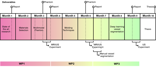
The project is divided in 3 work packages:
- Multimodal phantom development
- Patient specific phantom development
- Vessel segmentation analysis
Multimodal phantom development
Within this work-package initial research pertaining the state of the art of multimodal phantoms will be performed. This research is aimed at finding the materials which provides realistic US and MRI imaging. Research into see-through materials (for the parenchyma) will be also carried on, since this feature would enable surgeons to quickly evaluate the position of instrument with respect to internal structures.
The materials image properties will be evaluated by developing a simple phantom (containing parenchyma, vessels, and tumors) which will be scanned with US and MRI. The obtained images will be compared with real US and MRI scans, by analyzing the intensity values of the scans and the edges between different structures.
Patient-specific phantom development
Based on the results of WP1, a new patient specific phantom will be developed. The resulting phantom will have the same material properties to the one developed in WP1 with the addition of resembling a realistic liver.
This will be accomplished by developing a mold from a real 3D MRI scan. During this process, 3D printing techniques, as well as methods for accurately placing vessels and tumors within the phantom will be developed.
The shape of the patient-specific phantom will be evaluated by creating a 3D model of it through MRI scan. The resulting 3D model shape and volume, will be compared with the original 3D scan.
Vessels segmentation analysis
In order to accurately assess the vessels shape, deep learning segmentation techniques, aimed at reconstructing a 3D volume of the vessels, will be investigated.
Within this work package, 3D ultrasound scans of the phantom will be acquired. Initially, manual delineation of the imaged vessels will be performed. This manual delineation will result in a training dataset, which will be used to initialize automatic classification techniques.
Subsequently, different classification and feature extraction methods will be investigated and the resulting 3D model will be compared with the one obtained through manual segmentation.
Knowledge acquisition
During this project, the student will acquire knowledge in, MR and ultrasound image reconstruction and segmentation. The latter part will be developed by using deep learning algorithms which will allow to gain knowledge in medical image analysis and machine learning. Additionally 3D printing methods and casting techniques, will be also learned during this project.
Finally, scientific methodology and analysis will be learned through accuracy experiments and reports.
Prerequisites
- Enthusiastic Master student in electrical engineering, biomedical engineering, computer science, technical medicine or a related field
- A good team player with excellent communication skills
- A creative solution-finder
Duration: 40 weeks (M3)
Start date: a.s.a.p.
