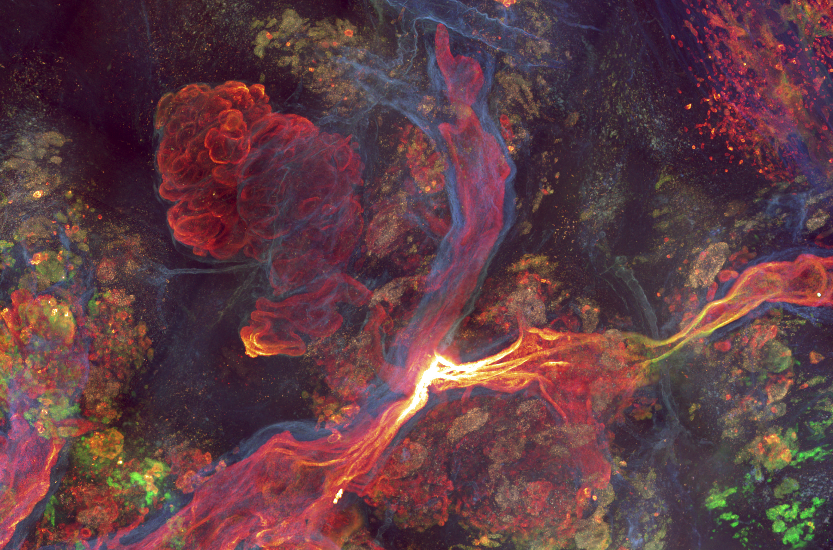
Our research has shown that in the mammary ductal epithelium, cellular hierarchy limits the clonal expansion of Brca1 and Tp53 mutant cells. However, repeated tissue remodelling during the hormonal cycle, through alveolar branching and regression, disrupts this constraint. Mutated cells may be lost during alveolar regression, but if they persist, they can drastically expand in the subsequent proliferative phase, generating large clonal fields that give rise to spontaneous tumors (Ciwinska, Messal and Hristova et al. Nature 2024).
Tumor-initiating cells distort epithelial geometry, modulate mechanical forces, and alter biochemical signals to generate permissive niches for expansion. We found that in pancreatic ducts, a KRAS^G12D-ERK-MEK-MLC2 axis induces cortical tension imbalances that direct lesion growth into the parenchyma or duct lumen depending only on the epithelial curvature. This early switch exposes lesions to distinct microenvironments, correlating with divergent invasive potential (Messal and Alt et al. Nature 2019).
These transformations occur across scales, from cytoskeletal changes in single cells to the reorganization of the local tissue architecture, and are essential for progression from mutation to malignancy. Our lab investigates these mechanisms through the lens of cancer microanatomy, integrating whole-organ tissue clearing and 3D microscopy with spatially guided molecular profiling and mechanistic in and ex vivo assays to understand how tumor-suppressive architecture is subverted into tumor-permissive landscapes.

