Support us

How can we use (bio)medical images to treat patients as accurately as possible?

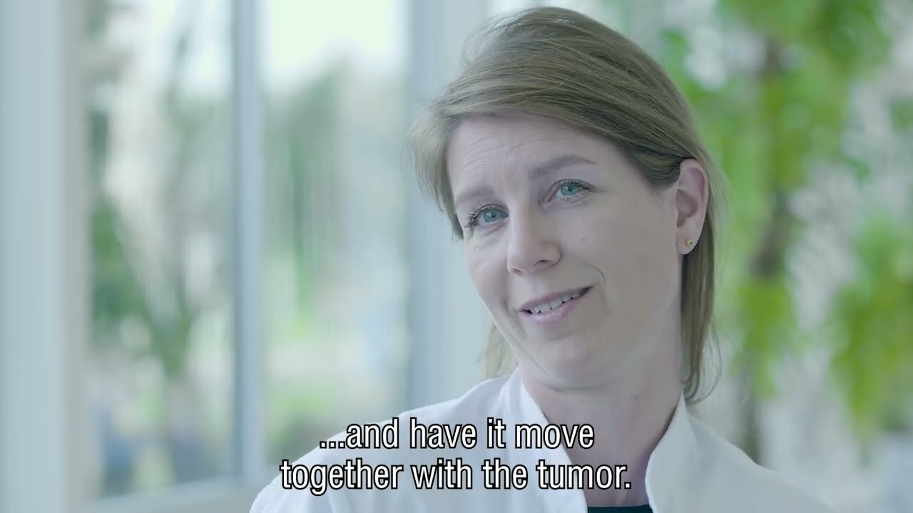
To operate and irradiate a tumor with great precision, surgeons and radiation oncologists need to know where exactly they can find the cancer cells. Physicians use several different medical scans that show them the tumor in relation to the rest of the body. But there are more possibilities. The location and shape of a tumor change in between treatments as well as during treatment like radiation or surgery. Tumors in the stomach and chest cavity move with every breath. And if a tumor shrinks as a result of treatment, or a patient loses weight, the radiation will need to be adapted. The Netherlands Cancer Institute researchers the various applications of medical imaging to better adapt treatment to the current situation of every individual patient.


Example projects

Liver irradiation

One of the things we are working on is the improvement of techniques to irradiate liver metastases. These tumors tend to move during treatment, which complicates things. They are not easy to spot on a CT scan either, which necessitates a marker in the tissue. Using a new irradiation device with a fully integrated MRI scanner (MR-linac), we no longer have to use a marker in the tissue. We can create a new radiation field right before every treatment session by making a scan to help adapt the treatment. We want to be able to create new scans during treatment and adapt the radiation field based on the movement we observe. By increasing our understanding of the precise location of the tumor, we can improve the aim of the radiation, which allows us to increase the dose without harming healthy tissue. 

Medical images to improve accurate treatment

We can make better use of medical imaging and use them more often to increase the accuracy of our treatments. Marlies Nowee explains her research on radiation techniques for liver metastases that move during the delivery of the treatment.

Hepatic surgery with ultrasound

We are investigating the possibility of using ultrasound technology to increase surgical precision. Surgeries and researchers use artificial intelligence to create a 3D model of the blood vessels and tumor in the operating room. The surgeon can use this to navigate their way around the liver during surgery while seeing an additional layer of information on top of the tissue: augmented reality.

This site uses cookies

This website uses cookies to ensure you get the best experience on our website.